Backup for http://www.popsci.com/node/36702.
| Capillary network M I Walker, Wellcome Images This striking image actually shows part of an ox's eye, and the capillaries in it. Capillaries are small blood vessels, which act as the connective network between arteries and veins. The capillaries have been made visible by injection of an insoluble dye into the artery that supplied them. |
|
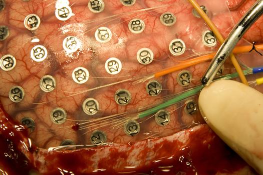
| Brain Array Courtesy Kelly Johnson/University of Utah Department of Neurosurgery Here, microwires emerging from the green and orange tubes connect to two arrays of 16 microelectrodes. Each array is embedded in a small mat of clear, rubbery silicone. The mats are barely visible in this image. These microelectrode arrays sit on the brain without penetrating it, a step toward longer-lived, less invasive versions of "neural interfaces" that in recent experiments elsewhere have allowed paralyzed people to control a computer cursor with their thoughts. The new microelectrode arrays were placed in two patients at the University of Utah who already were undergoing brain surgery for severe epilepsy. The larger, numbered, metallic electrodes are used to locate the source of epileptic seizures in the brain, so the patients allowed the microelectrodes to be placed on their brains at the same time. |
|
| Hairy Hearing Courtesy SPL / Photo Researchers, Inc. Inner ear hair cells. Colored scanning electron micrograph (SEM) of sensory hair cells from the organ of corti, in the cochlea of the inner ear. These cells are surrounded by a fluid called the endolymph. As sound enters the ear it causes waves to form in the endolymph, which in turn cause these hairs to move. The movement is converted into an electrical signal, which is passed to the brain. The V-shaped arrangement of hairs lies on the top of a single cell. Magnification: x21,000 when printed 10cm wide. | 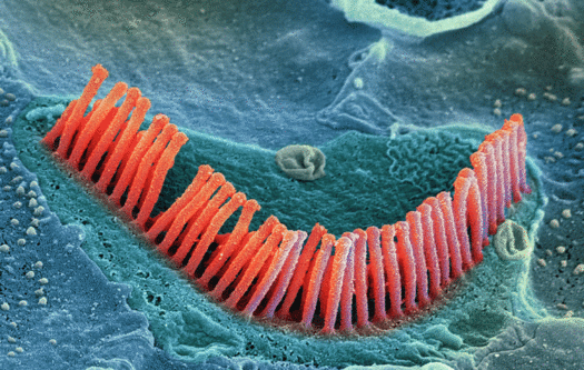 |
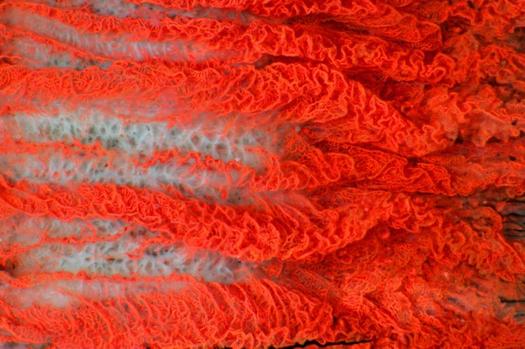
| Anglerfish Ovary, Magnified 4x Courtesy Nikon Small World Photo Micrography Competition: Photographed by James Hayden This cross-section of an anglerfish’s ovary is reminiscent of another sea creature—the nautilus. Its otherwise-dull spirally composition caught James Hayden’s eye, so he used fluorescent hematoxylin and Eosin stains to illuminate the different details of the image. “I was trying to create an image that sharply defined the boundaries of the different parts of the specimen, so that the image could actually be used to demonstrate the morphology of the ovary and eggs,” he says. | 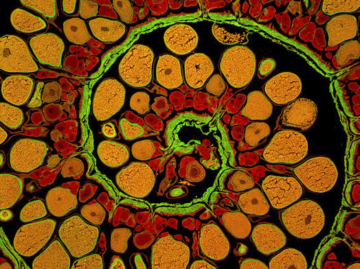 |
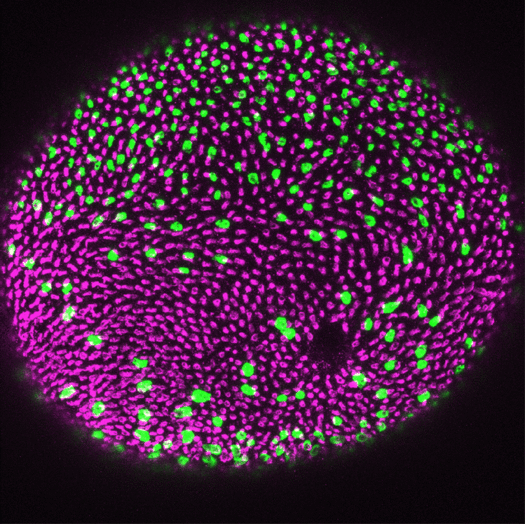
| Zebrafish Retina Courtesy Associate Professor James Fadool and Karen Alvarez-Delfin/Florida State University This fireworks display is actually a microscope image of a zebrafish retina that's been immunolabeled for ultraviolet cones (magenta) and rods (green). The image shows the regular pattern of the cones and the scattered pattern of the rods typical of a normal fish. The labeling was performed by Karen Alvarez-Delfin, doctoral candidate at Florida State University. |
|